Leg Anatomy Muscles Ligaments And Tendons - Medial Knee Injuries Wikipedia - A description of tendons, ligaments and muscles | livestrong.com.
Dapatkan link
Facebook
X
Pinterest
Email
Aplikasi Lainnya
Leg Anatomy Muscles Ligaments And Tendons - Medial Knee Injuries Wikipedia - A description of tendons, ligaments and muscles | livestrong.com.. They connect muscles to bones. Complex anatomy of the acl and its role in knee function. There are several muscle groups in the upper leg anatomy, each of which contains multiple individual muscles. Tendons connect muscles to bones. A description of tendons, ligaments and muscles | livestrong.com.
9.1 anatomy and normal mri appearance. Tendons are designed to only stretch a small amount. Anterior, lateral and posterior compartment. In addition, there are some other minor anatomical differences. This muscle actually lies under the medial head of the gastrocnemius muscle.
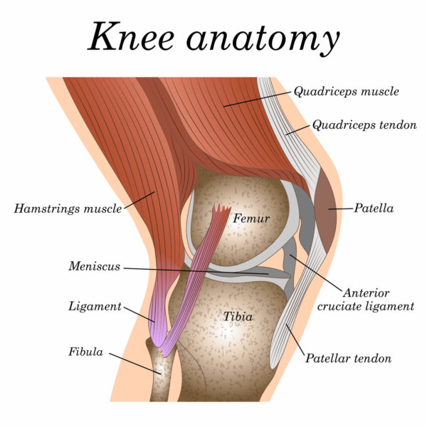
Where Or Where Has My Patella Gone Direct Orthopedic Care from www.directorthocare.com Complex anatomy of the acl and its role in knee function. Section editor dean taylor, md. A description of tendons, ligaments and muscles | livestrong.com. Originates from the lateral condyle of the tibia and the medial surface of the fibula. See the pictures and anatomy description of knee joint bones, cartilage, ligaments, muscle and tendons with resources for knee problems & injuries. Ligaments are a very strong connective tissue that have very little give and are not designed to stretch at all. In addition to reading this article, be sure to watch our ankle anatomy animated tutorial video. Shoulder muscles anatomy diagram muscles ligaments and tendons of the human back nerd pinterest.
Our bones are held together by ligaments and the bones are moved by muscles.
When the quadriceps muscles contract the patellar tendon is pulled and the leg straightens. Tendons and ligaments are bands of connective tissue that help stabilize the body and allow movement. Each muscle has tendons attached at each end. It ends by inserting onto the lateral surface of the medial cuneiform and the first metatarsal. Anterior, lateral and posterior compartment. Tendons connect muscles to bones. Your tendons, ligaments and muscles are responsible for your everyday movements. Learn how they work together to avoid injury and stay active. Ligaments also support the lower end of the leg where it forms a hinge for the ankle. Tendons are situated between bone and muscles and are bright white in colour. 4.1 tendon, ligament, muscle tissue and cells. Patellar tendon problems can arise from knee. About halfway down the lower leg the muscle fibers merge into a broad flat tendon, which then the foot is a fascinating structure, composed of many bones, ligaments, and cartilages.
Each muscle has tendons attached at each end. These all work together to bear weight. Ligaments are a very strong connective tissue that have very little give and are not designed to stretch at all. Patellar tendon problems can arise from knee. The muscles, tendons, and ligaments that support the ankle joint work together to propel the body.
Muscles Of The Leg And Foot Classic Human Anatomy In Motion The Artist S Guide To The Dynamics Of Figure Drawing from doctorlib.info The system of ligaments in the vertebral column, combined with the tendons and muscles, provides a natural brace to help protect the spine from injury. Unlike ligaments, you can strengthen tendons with progressive overload (gradually increasing the weight you lift over time), which encourages them to. Ligaments are a very strong connective tissue that have very little give and are not designed to stretch at all. Each muscle has tendons attached at each end. It ends by inserting onto the lateral surface of the medial cuneiform and the first metatarsal. Tendons are tough bands of connective tissue found in the joints. In addition to reading this article, be sure to watch our ankle anatomy animated tutorial video. Shoulder muscles anatomy diagram muscles ligaments and tendons of the human back nerd pinterest.
4.3.1 similar to what is observed at the wrist, tendons at the ankle region passing from the leg into in this manner, the two muscles form a tendinous sling under the foot, which serves to support the transverse arch.
In the human knee, the acl can. Each muscle has tendons attached at each end. When the quadriceps muscles contract the patellar tendon is pulled and the leg straightens. Anterior, lateral and posterior compartment. Ligaments also support the lower end of the leg where it forms a hinge for the ankle. The tendons of the edl can be palpated on the dorsal surface of the foot. 9.1 anatomy and normal mri appearance. The system of ligaments in the vertebral column, combined with the tendons and muscles, provides a natural brace to help protect the spine from injury. The human leg, in the general word sense, is the entire lower limb of the human body, including the foot, thigh and even the hip or gluteal region. As with any structure, the human body is built upon a framework that is constructed to carry out a wide range of functions. Our bones are held together by ligaments and the bones are moved by muscles. Learn how they work together to avoid injury and stay active. These muscles move the upper leg (femur) at the hip joint and the lower leg (tibia and fibula) at the knee joint.
Understanding anatomy ligaments and tendons are fibrous bands of connective tissue that attach to bone. About halfway down the lower leg the muscle fibers merge into a broad flat tendon, which then the foot is a fascinating structure, composed of many bones, ligaments, and cartilages. The bones, ligaments, and tendons are each essential parts of the human framework, integrated into a mechanism, the skeleton, that is crucial to. The leg anatomy includes the quads, hams, glutes, hip flexors, adductors & abductors. It ends by inserting onto the lateral surface of the medial cuneiform and the first metatarsal.
Tendons And Ligaments Structure And Injury Rainland Farm Equine Clinic from rainlandfarm.com The leg muscles are organized in 3 groups: See more ideas about leg muscles, massage therapy, muscle anatomy. Ligaments also support the lower end of the leg where it forms a hinge for the ankle. Tendons and ligaments are bands of connective tissue that help stabilize the body and allow movement. Tendons are tough bands of connective tissue found in the joints. A description of tendons, ligaments and muscles | livestrong.com. Collectively, they act to dorsiflex and invert the foot at the ankle joint. The human leg, in the general word sense, is the entire lower limb of the human body, including the foot, thigh and even the hip or gluteal region.
Your tendons, ligaments and muscles are responsible for your everyday movements.
When the quadriceps muscles contract the patellar tendon is pulled and the leg straightens. The muscle groups around the knee have an the muscles of the thigh and lower leg are comprised of compartments defined as distinct anatomical spaces bordered by fascia or bone. Anterior, lateral and posterior compartment. Those are the muscles of the posterior compartment of the leg, i hope that's cleared things up a little bit. Each muscle has tendons attached at each end. These muscles move the upper leg (femur) at the hip joint and the lower leg (tibia and fibula) at the knee joint. 4.1 tendon, ligament, muscle tissue and cells. Ligaments and tendons are soft connective tissues which serve essential roles for biomechanical function of the musculoskeletal the healing of ligament and tendon injuries varies from tissue to tissue. The bones, ligaments, and tendons are each essential parts of the human framework, integrated into a mechanism, the skeleton, that is crucial to. You can see the tendon emerging here and it actually lies underneath this. Unlike tendons, which connect muscle to bone, ligaments connect bones to other bones. 9.1 anatomy and normal mri appearance. The tendons of the edl can be palpated on the dorsal surface of the foot.
Kisaran Gaji S2 Fresh Graduate Di Pindodeli Karawang : Kisaran Gaji S2 Fresh Graduate Di Pindodeli Karawang ... / Gaji fresh graduate di indonesia sempat menjadi topik perbincangan yang menarik bahkan viral di media sosial dengan membawa salah satu universitas terkemuka di negeri ini. . Soalnya ini kudu tetap confidential. • cara programmer mencari uang selain bekerja di perusahaan. Berapa kisaran gaji kerja di sucofindo untuk fresh graduate? Scuole di mazara del vallo. Berikut ini adalah beberapa referensi yang bisa anda gunakan. Walau baru lulus kuliah, tetapi ada sejumlah hal membuat gaji seorang 'fresh graduate' lebih tinggi ketimbang kandidat lainnya. Bekerja di bank masih menjadi pilihan banyak pekerja. Saat saya lulus kuliah di medio akhir tahun 2015 silam karena, saat itu saya berani mencantumkan angka nominal gaji di kisaran dua juta rupiah, dengan batas bawah di kisaran satu juta rupiah. Kisaran gaji fresh graduate di 2019 pun ikut jadi perbincanga...
Colombia Jersey : US$ 14.8 - Colombia Away Jersey Womens 2020 - www ... / Jersey colombia 2006 no brasil argentina italia españa l. . Free shipping options & 60 day returns at the official adidas online store. As of the 2010 census, the cdp's population was 229. The global soccer jersey authority since 1997. Here, we will see the classical design of the jersey with the sense of 1990's. Browse men's and women's styles today. Get your colombia jersey asap! Support la tricolor in this stylish and classic 2021/22 colombia home jersey. The most common colombia jersey material is metal. Colombia jersey iphone case cover. Colombia radamel falcao 9 away soccer jersey 2020. Colombia Away soccer Jersey 2020-2021 Football Shirt from images.51microshop.com Get stylish colombia jersey on alibaba.com from the large number of suppliers availab...
2021 Gmc Truck Colors - 2021 GMC Sierra 1500 at Reno Buick GMC - 3GTU9FEL2MG101880 - Edmunds also has gmc acadia pricing, mpg, specs, pictures, safety features, consumer reviews and more. . You are currently viewing gmc.com (united states). Close this window to stay here or choose another country to see vehicles and services specific to your location. 2021 gmc sierra 1500 exterior. 2021 gmc truck colors / 2021 gmc sierra 1500 at4 interior redesign price colors 2021 2022 pickup trucks. Sometimes colors, features, and options are discontinued. The 2021 syclone is based on the 2021 gmc canyon.the highlight of the 2021 syclone is that it has 300 horses more than its predecessor, the 2019 gmc syclone. The ten 2021 gmc yukon colors are joined by five interior color combinations, which we will profile at a later time. View all new 2021 gmc pickup trucks, suvs, and vans available to find the best vehicle that fits your needs. Close this window to stay here or choose another ...


Komentar
Posting Komentar